 FHL’s “core courses” (which are partially supported by our Marine Life Endowment) include Invertebrate Zoology, Marine Botany, Fishes, and Developmental Biology – each of which is taught in various forms, with different instructors and foci. Taking one of these courses often launches students into careers as they find an unexpected passion. Our fish morphology-related courses, REU internships, and other programs have a long and rich history of training generations of vertebrate functional morphologists and biomechanists like Emily Carr. We love to see these students out in the world with excellent jobs and producing wonderful publications!
Best,
Dr. Megan Dethier, FHL Director
Make your gift today
Illuminating the Past: From FHL to Fluorescent Fish
by Emily Carr

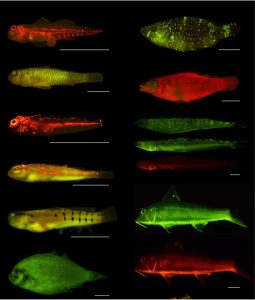
Life for fish on coral reefs is very chaotic. It’s almost like Times Square: lots of bright colors, flashy lights, and countless different organisms swimming every which way. In this chromatic chaos, any way to stand out to members of your own species may be advantageous. One way scientists think fish may be able to stand out is through biofluorescence (Figure 1). Biofluorescence is a passive process where sunlight is absorbed by tissues and re-emitted at longer wavelengths (e.g. green, orange, red). This phenomenon is widespread across the tree of life, including groups like salamanders, mammals, and fish (Carr et al. 2025, Marshall and Johnson, 2017). However, biofluorescence is particularly diverse in shallow water reef fish, and we think it may be because of the visually chaotic environment of coral reefs (Carr et al. 2025). I’m currently studying biofluorescence at the American Museum of Natural History, but my interest in a research-based career began, like many, at Friday Harbor Labs (FHL).
During my undergraduate degree, I visited FHL for three summers (I couldn’t get enough!). During the first summer, I worked with Karly Cohen, a then-PhD student, to model how forces (e.g. biting prey) affected different shapes of CT-scanned fish teeth. Though I spent most of my time cooped up in a back room desperately trying to learn how to use 3D modeling software, I fell in love with FHL. It was a whole new world of creative, fun, and at times crazy scientists studying organisms in all kinds of unique ways.

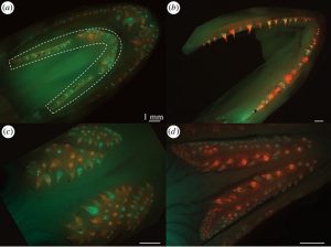
The following summer, when I heard about the 2020 Fish Class (Functional Morphology and Ecology of Marine Fishes), I was all in (despite the global pandemic happening at the time). While masked up and six feet apart, I began another project with Karly Cohen and Dr. Adam Summers measuring tooth replacement rates of Pacific lingcod (Carr et al. 2021). We used two fluorescent dyes to determine which teeth were older (red) versus newly developed and replaced (green) over a period of two weeks (Figure 2). I then counted over 10,000 tiny fluorescent teeth under a microscope in a dark room – the perfect job for an overzealous undergrad! Thankfully, all my hard work paid off. We found a remarkably fast rate of tooth development: 20 teeth per day of the ~500 total teeth in Pacific lingcod. In humans, that would be like losing and replacing one tooth every single day! Be thankful you’re not a lingcod.
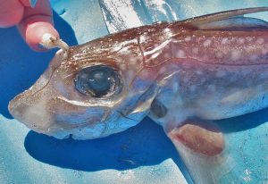
As a now self-proclaimed fish dentist, I began looking for new research opportunities related to tooth development. In a greatly desired turn of events, I came back to FHL the next summer to do just that, this time as a student in the NSF-funded REU program (Research Experience for Undergraduates). I worked with Dr. Gareth Fraser on a project investigating an odd structure called a tenaculum in the spotted ratfish, a species of chimaera (Figure 3). Chimaeras are cartilaginous fish closely related to sharks, skates, and rays. The tenaculum is a secondary copulatory structure on the forehead of male chimaeras. The males are thought to use this toothy tenaculum to grab onto females during mating (Carrier et al. 2022). Also be thankful you’re not a female chimaera!

During the REU program, we took samples of the tenaculum for histology, a method of differentially dyeing tissues to determine tissue types. We thinly cut the tenaculum with the scientific equivalent of a very precise deli meat slicer and put the tissue through a series of dyes. We found that the teeth of the tenaculum develop more similarly to shark teeth than they do to the rabbit-like teeth in the mouths of chimaeras. This was an interesting finding, because it revealed a potential decoupling of the tooth development systems between the mouth and tenaculum of male chimaeras, despite their proximity on the head.
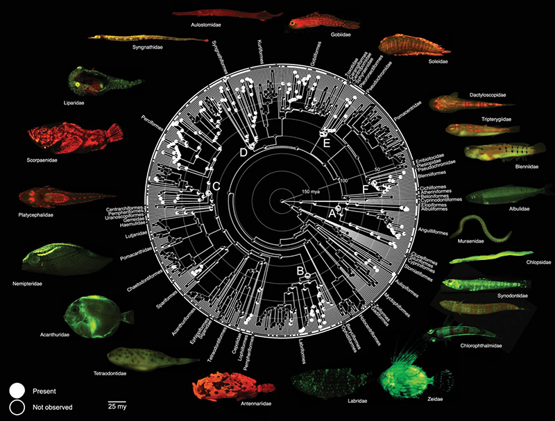
During that summer at FHL, Dr. Adam Summers told me about a potential PhD program that fit my interests; I later found myself joining that program after I graduated. I am now a PhD student at the American Museum of Natural History’s (AMNH) Richard Gilder Graduate School in New York. My dissertation is focused on light phenomena, including both biofluorescence and bioluminescence, and diversification in fishes. While biofluorescence is a passive process of shifting wavelengths of light, bioluminescence is the creation of light by an organism, most often seen in the deep sea. Biofluorescence relies on external light to function, so it is restricted to more shallow water environments with sufficient ambient light. Recently, we conducted a large-scale study of biofluorescent fish to try to understand the origin, evolutionary patterns, and trends in biofluorescence across a family tree (Carr et al. 2025, Figure 4). We found that biofluorescence first evolved ~112 million years ago in eels, which have both red and green fluorescence. We also found that reef-associated species are much more likely to evolve biofluorescence than fishes in other habitats. The close association between biofluorescence and reef environments is likely because of the complex chromatic conditions of coral reefs, like the Times Square of the ocean.

I conducted this research at the AMNH, but it all truly started at FHL. While I am now on the opposite coast studying different things, the experiences and research I was exposed to at FHL led me to where I am today. I’m eternally grateful for the funding, mentors, and fellow students who have supported me along the way.
Funding:
Adopt A Student Fund, University of Washington, Friday Harbor Labs
Kozloff Undergrad Endowment Fund, University of Washington, Friday Harbor Labs
NSF Research Experience for Undergraduates, University of Washington, Friday Harbor Labs
Graduate Research Fellowship Program, National Science Foundation
Ph.D. Student Fellowship, Richard Gilder Graduate School, American Museum of Natural History
References:
Carr E.M., Thurman M.A., Martin R.P., Sparks T.S. and J.S. Sparks. 2025. Marine fishes exhibit exceptional variation in biofluorescent emission spectra. PLOS ONE: 20(6). doi:e0316789.
Carr E.M., Martin R.P., Thurman M.A., Cohen K.E., Huie J.M., Gruber D.F., et al. 2025. Repeated and widespread evolution of biofluorescence in marine fishes. Nat Commun.: 16(1), 4826.
Carr E.M., Summers A.P. and K.E. Cohen. 2021. The moment of tooth: rate, fate and pattern of Pacific lingcod dentition revealed by pulse-chase. Proc R Soc B Biol Sci.: 288(1960), 20211436.
Carrier J.C., Simpfendorfer C.A., Heithaus M.R. and K.E. Yopak. 2022. Biology of Sharks and Their Relatives. New York: CRC Press.
Marshall J. and S. Johnsen. 2017. Fluorescence as a means of colour signal enhancement. Philos. Sci.: 372. doi:20160335.
Tide Bites is a monthly email with the latest news and stories about Friday Harbor Labs. Want more? Subscribe to Tide Bites or browse the archives.