One of (many) hard things about the COVID-19 pandemic, for the whole world, was the drastic curtailing of human interactions. We felt this at FHL, of course, but also suffered from the fact that resident and visiting scientists were restricted from spontaneous collaborations like the one discussed in this month’s Tide Bite. When seminars are on Zoom, most participants can’t chat afterwards to compare interests and generate excitement about new scientific connections. Some great interactions have sprung from shared lunches in the Dining Hall, chance meetings at a common-use piece of equipment, or spontaneous chats while collecting organisms off the breakwater. FHL is indeed a “real-world petri dish” where unexpected things grow!
Some of Olivia’s previous, foundational coursework and much of her graduate research was supported by generous donors to FHL (p.2 of this newsletter). Private donations provide essential funding for FHL students and scientific research. The Adopt-a-Student Fund and the Research Fellowship Endowment are both excellent investments in our future!
Incubating Real-World Connections at Friday Harbor Labs
by Olivia Graham and Eric Edsinger
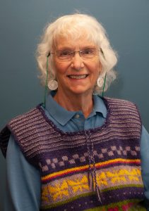
Olivia Graham is a postdoc with Dr. Drew Harvell at Cornell University and has been studying eelgrass at FHL for 9 years. She loves connecting with other researchers at the Labs.
Eric Edsinger is a research scientist at the University of Florida Whitney Laboratory for Marine Biosciences and works at the intersection of biodiversity, imaging, and sequencing. He loves walking the docks at FHL to see what species are out in the waters, drifting by in the currents.

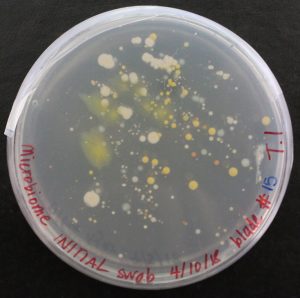
“How’s everybody doing today? Growing nice, big, strong, and infectious?” I ask brightly, opening the door to my humming incubator. I am checking on cultures of Labyrinthula zosterae (Lz), a marine protist that infects eelgrass (Zostera marina), the most common cold-water seagrass species worldwide. Not only am I growing Lz, but also the bacteria that live on the surface of these marine plants. Peering at one of the dishes, I happily see a kaleidoscope of cultures: fuzzy yellow ones, tiny tangerine ones, and splotchy, white ones. Marveling at the vibrant community growing on this little dish, I wonder what type of interactions these microbes might have with one another and Lz.
Understanding the role of the eelgrass microbiome (bacteria living on the surface of eelgrass) in Lz defense was one of the primary questions I explored in my PhD dissertation, and is a key area of research our team is currently pursuing, led by new FHL postdoc Dr. Rebecca Maher (stay tuned for more research updates in a future TideBite). For now, I scrape some of the sticky cells off of an agar petri dish, onto a hemocytometer (specialized glass slide for counting cells), and carefully place it onto the loading platform of my microscope. “Am I looking at the right thing? Or are those just bubbles? Heck, am I even still looking at the glass slide??” As I slowly turn a knob and mutter to myself, the shapes become more distinct, the edges more clearly defined. Smiling, I can now see the spindle-shaped Lz cells, clearly illuminated and magnified. Visualizing Lz, of course, enables me to carefully study this pathogen and see patterns that may not be recognizable otherwise, not unlike satellite images of Earth that provide us with new perspectives. Peering through the microscope, I watch the cells glide along their slimy networks. As a cousin to slime molds, Lz secretes mucus tracks that allow the cells to move across the agar – or, in nature – through eelgrass cells.

Though small, Lz can have big ecological impacts on foundational eelgrass meadows. Eelgrass provides essential ecosystem services to coastal areas, creating key habitats for juvenile fish, filtering pathogens from seawater, and efficiently sequestering carbon belowground. Despite these superpowers, eelgrass is not immune to stressors. Lz is the causative agent of seagrass wasting disease (SWD), which decimated eelgrass meadows along the Atlantic Coast of the USA and Europe in the 1930s and 1940s. Disease outbreaks continue today, particularly in the San Juan Islands, WA (Groner et al. 2021, Aoki et al. 2022, 2023, Graham et al. 2023; 2019 TideBite). Field experiments on San Juan Island, WA also show that Lz can reduce eelgrass growth and belowground sugar reserves, upon which these valuable plants rely to survive during harsh winters (Graham et al. 2021).
As a marine scientist studying seagrass wasting disease, I consider myself lucky because I work with a pathogen that is known and culturable. For many marine diseases – such as sea star wasting disease – the causative agent remains unknown and many other pathogens are not culturable. Having a known, culturable pathogen allows our lab to do controlled inoculation experiments and keep “pet” cultures of the pathogen in an incubator. Very convenient!

Of course, fieldwork is my scientific bread and butter. While I certainly use robust laboratory techniques, my preferred approaches for tackling thorny questions about marine disease dynamics include field surveys and experiments that employ snorkeling, SCUBA, and a trusty clipboard and pencil. Though this skill set serves me well most of the time, I have never been able to precisely image Lz. My feeble attempts at photographing the slimy cells by steadying my cell phone camera against microscope eyepieces have produced lackluster photos. Likewise, my photos of the milky culture on agar plates are unlikely to make the cover of Environmental Microbiology. Fortunately, meeting Dr. Eric Edsinger changed all of that.

Eric first met Dr. Drew Harvell, Professor Emerita at Cornell University and Affiliate Faculty at UW SAFS and FHL researcher, when Eric was last at the Labs almost two decades ago. He returned to FHL this last year for research and teaching. Eric and Drew reconnected after giving short talks on their work to the FHL community. Upon learning that Drew’s team works with Lz, a culturable marine protist, Eric eagerly offered to send our Lz cultures for genetic sequencing. “Amazing! Could you also work your magic with the confocal microscope to image Lz inside eelgrass or zoospores in Lz cultures?” Drew wondered. She hoped that by experimenting with staining and imaging techniques, Eric might be able to capture clear photos of the small, sticky, spindle-shaped cells. Once we gave Eric a few petri dishes with Lz cultures and bought a few snazzy stains for him to test, he set to work.
Thanks to Eric’s hard work we now have terrific images of Lz, which help illuminate this important pathogen in presentations and reports to technical and general audiences alike. Working with a microscopic pathogen means that when giving presentations to students, previously I could only show them agar dishes covered in the milky cultures. Eric’s sharp images now bring Lz into better focus for me and those with whom I share my work.

In this way, FHL is a petri dish: it brings together scientists from all walks of life and career stages and after some incubation period, there is suddenly a robust, diverse community with all sorts of exciting interactions. Not unlike my agar dishes with vibrant and funky microscopic critters growing on them, the Labs serve as an incredible facilitator for unique interactions amongst scientists of all ages. Normally, I would not have worked closely with an imaging specialist like Eric. Yet, by both being at FHL, we could collaborate on a fun, rewarding side project. This is a prime example of Friday Harbor Labs’ forte: bringing together scientists and creating meaningful connections. (And as an aside, I can attribute much of my research career path to similar “petri dish” interactions at FHL!) Of course, specialized equipment at the Labs enables some of these collaborations, but there is also an inherent je ne sais quoi about FHL that allows these interactions to truly flourish. If you haven’t already, I sincerely hope you can experience this petri dish magic sometime.
References
Aoki L.R., Yang B., Graham O.J., Gomes C., Rappazzo B., Hawthorne T.L., Duffy J.E., and D. Harvell. 2023. UAV high-resolution imaging and disease surveys combine to quantify climate-related decline in seagrass meadows. In Frontiers in Ocean Observing: Emerging Technologies for Understanding and Managing a Changing Ocean. Kappel E.S. et al. eds, Oceanography 36 (Supplement 1). https://doi.org/10.5670/oceanog.2023.s1.12.
Aoki L.R., Rappazzo B., Beatty D.S., Domke L.K., Eckert G.L., Eisenlord M.E., et al. 2022. Disease surveillance by artificial intelligence links eelgrass wasting disease to ocean warming across latitudes. Limnology & Oceanography: 67, 1577–1589. doi: 10.1002/lno.12152.
Graham O.J., Aoki L.R., Stephens T., Stokes J., Dayal S., Rappazzo B., et al. 2021. Effects of Seagrass Wasting Disease on Eelgrass Growth and Belowground Sugar in Natural Meadows. Front. Mar. Sci.: 8(768668).
doi: 10.3389/fmars.2021.768668.
Graham O.J., Stephens T., Rappazzo B., Klohmann C., Dayal S., Adamczyk E.M., et al. 2023. Deeper habitats and cooler temperatures moderate a climate-driven seagrass disease. Phil. Trans. R. Soc. B: 378(20220016).
doi: 10.1098/rstb.2022.0016.
Groner M., Eisenlord M., Yoshioka R., Fiorenza E., Dawkins P., Graham O., et al. 2021. Warming sea surface temperatures fuel summer epidemics of eelgrass wasting disease. Mar. Ecol. Prog. Ser.: 679, 47–58.
doi: 10.3354/meps13902.
Tide Bites is a monthly email with the latest news and stories about Friday Harbor Labs. Want more? Subscribe to Tide Bites or browse the archives.
